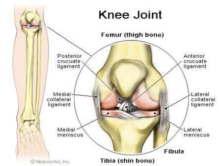
Knee Joint
The knee is the largest synovial joint in the body. It is composed of 3 bones and 3 joints although 2 of the 3 joints share a common cavity.
The bones of the knee comprise the femur (thigh bone), tibia (shin bone), patella (kneecap), and to a lesser degree the fibula. The knee joint itself is made up of the tibio-femoral joint, which itself is comprised of a medial compartment and a lateral compartment. The true knee joint also includes the patello-femoral joint. Another important component of the knee joint complex, although not part of the true knee joint, is the superior tibio-fibula joint.

The femur is the longest and strongest bone in the body. The shaft of the femur is nearly cylindrical, fairly uniform in calibre and the shaft has a bow with the apex anteriorily progressing from proximal to distal.
The distal aspect of the femur broadens into the medial and lateral condyles with all but the sides of these condyles being articular and involved in the knee joint. The inferior, posterior oblong portions of the condyles articulate smoothly with the tibial plateau, whereas the central anterior surface between the condyles articulates with the facets of the patella. The inferior, oblong surfaces of the condyles are separated by the intercondylar fossa which houses the cruciate ligaments.
In shape and dimensions the femoral condyles are asymmetric; the larger medial condyle has a more symmetrical curvature. The lateral condyle viewed from the side has a sharply increasing radius of curvature posteriorily. The lateral condyle is slightly shorter than the medial. The long axis of the lateral condyle is slightly longer and is placed more sagittal than the long axis of the medial condyle. The lateral condyle is slightly wider than the medial condyle at the centre of the intercondylar notch. Anteriorily the condyles are separated by a groove, the femoral trochlea. The sulcus represents the deepest point in the trochlea relative the mid plane between the condyles, the sulcus lies slightly laterally. The lateral condyle has a greater posterior excursion than the medial. The contact surface of the patella is derived mostly from the lateral condyle. The anterior extensions of both condyles form a fossa for the patella to sit in extension. The lateral extension is the greatest.
The intercondylar notch separates the two condyles distally and posteriorily. The lateral wall of the notch has a flat impression where the proximal origin of the anterior cruciate ligament (ACL) arises. On the medial wall of the notch is a larger site where the posterior cruciate ligament (PCL) originates.
The lateral condyle has a short groove just proximal to the articular margin, in which lies the tendinous origin of the popliteus muscle. The groove separates the lateral epicondyle from the joint line. The lateral epicondyle is a small but distinct prominence which attaches the lateral (fibula) collateral ligament (LCL). On the medial condyle the prominent adductor tubercle is the insertion of the site of the adductor magnus. The medial epicondyle lies anterior and distal to the adductor tubercle and is C shaped with a central depression or sulcus. The epicondylar axis passes through the centre of the sulcus of the medial epicondyle and the prominence of the lateral epicondyle. This line serves as an important reference line in total knee replacements. The medial epicondyle is more prominent and provides attachment for the medial (tibial) collateral ligament (MCL).
 The fibula is a long slender bone that lies parallel and lateral to the tibia. It does not participate in weight-bearing but rather serves the muscle and tendon attachments. The head of the fibula is knob-like, and superiorly slanted towards the tibia, is the almost circular articular surface which participates in tibiofibular articulation. At the postero-lateral limit of the articular facet the apex of the head projects upwards and provides attachment for the lateral (fibula) collateral ligament (LCL) of the knee joint. The tendon of biceps femoris muscle attaches to the lateral aspect of the head of the fibula.
The proximal tibia is expanded to receive the condyles of the femur. The shaft of the bone flares out into lateral or medial buttresses which form the medial and lateral condyles. The tibia is the weight bearing bone of the leg whereas the fibula serves the muscular attachments and for completion of the ankle joint. The superior articular surface of the tibia presents two facets. The larger medial facet is oval in shape and has a slight concavity. The lateral facet is nearly round and although concave from side to side, it is convex in front. The rims of the facets are in contact with the medial and lateral menisci but the central portions receive the condyles of the femur. Both tibial facets have a posterior inclination with respect of the shaft of the tibia of approximately 10°. On inspection of the tibial plateau, it would appear as though the femoral and tibial surfaces do not conform. However, this is more apparent than real. In the intact knee the menisci enlarge the contact area considerably and increase conformity of the joint surfaces.
The medial portion of the tibia between the articular facets is occupied by an intercondylar eminence with two tubercles. The articular surface of the tibia continues onto the adjacent sides of the medial and lateral intercondylar eminences. Anterior to the intercondylar eminence is a depression, the anterior intercondylar fossa to which, from anterior to posterior, the anterior horn of the medial meniscus, the ACL and the anterior horn of the lateral meniscus are attached. Behind this region are two elevations, the medial and lateral tubercles. They are divided by a gutter-like depression, the intertubecular sulcus. On an antero-posterior radiograph the medial tubercle usually projects more superior than the lateral tubercle. On the lateral radiograph the medial tubercle is located anterior to the lateral tubercle. The tubercles do not function as attachment sites for the cruciate ligaments or the menisci but may act as side to side stabilisers by projecting towards the inner sides of the femoral condyles. In the posterior intercondylar fossa, behind the tubercles, the lateral then the medial menisci are attached anterior to posterior. Most posterior, the posterior cruciate ligaments inserts on the margin of the tibia between the condyles. From its origin, the posterior cruciate ligament travels anterior and slightly medial where it is joined by one or two cords from the lateral meniscus (the anterior and posterior menisco-femoral ligaments (or the ligament of Humphrey and the ligament of Riesburg respectively) to attach the medial condyle of the femur in the intercondylar notch.
On the anterior aspect of the proximal tibia, the tibial tuberosity is the most prominent feature and is the attachment site of the patellar tendon. Approximately 2 to 3 cm lateral to the tibial tubercle is Gurdy’s tubercle, which is the insertion site of the ilio-tibial band (ITB). Posterior to Gurdy’s tubercle, the lateral condyle has a nearly circular facet on its postero-inferior surface for articulation with the head of the fibula.
At its upper medial portion the tibia provides the attachment for the medial collateral ligament, both the deep and superficial bands of the medial collateral ligament. The medial surface of the body of the tibia is smooth and convex. Its upper one third receives the insertion of the sartorius, gracilis and semitendinosus tendons (medial hamstrings).
The patella is the largest sesamoid bone in the body and is situated in the tendon of the quadriceps femoris muscle. It articulates against the anterior articular surface of the distal femur. It holds the patellar tendon off the distal femur thus improving the angle of approach of the tendon to its distal insertion on the tibial tuberosity, so increasing the power generation of the quadriceps mechanism by 30%. The anterior surface of the patella is convex. The superior border is thick and gives attachment to the tendinous fibres of the rectus femoris and vastus intermedius muscles. The lateral and medial borders are thinner and receive the tendinous fibres of the vastus lateralis and vastus medialis muscles respectively. These two borders converge inferiorly to the pointed lower pole of the patella which gives attachment to the patellar ligament.
The articulation between the patella and femoral trochlea forms the patello-femoral knee joint compartment. The articular surface of the patella is described as possessing seven facets. Both the medial and lateral facets are divided vertically into approximately equal thirds, whereas a seventh or odd facet lies along the extreme medial border of the patella. Overall the medial facet is smaller and slightly convex, the lateral facet which consists of roughly two thirds of the patella has both sagittal convexity and coronal concavity.
Six variants of the patella have been described. Types one and two are stable whereas the other variants are more likely to result in lateral subluxation as a result of unbalanced forces. The facets of the patella are covered by the thickest hyaline cartilage in the body which may measure up to 6.5mm in thickness.
The patella fits in the trochlea of the femur imperfectly with the contact area between the patella and femur varying with position of flexion. The area of contact never exceeds about one third of the total patella articular surface. At 10 to 20° of flexion at the distal pole the patella first contacts the trochlea in a narrow band across the medial and lateral facet. As flexion increases the contact area moves proximally and laterally. The most extensive contact is made at about 45° where the contact area is an elipse across the central portion of the medial and lateral facet. By 90° the contact area is shifted to the upper part of the medial and lateral facets. With further flexion the contact area separates into two distinct medial and lateral patches. Because the odd facet only makes contact with the femur in extreme flexion (such as in the act of squatting) this facet is habitually a non contact zone in humans in Western countries.
The main biomechanical function of the patella is to increase the momentum of the quadriceps mechanism. The load across the joint rises as flexion increases but because the contact area also increases, higher forces are dissipated over a larger area. However, if extension against resistance is performed, the force increases while the contact area shrinks. Straight leg raises eliminate forced transmission across the patello-femoral joint because in full extension the patella has not yet engaged the trochlea.
The joint between the circular facet of the head of the fibula and a similarly shaped surface on the postero-lateral aspect of the under surface of the lateral condyle of the tibia is a plane joint. The articular surface of the head of the fibula is directed superiorly and slightly antero-medially to articulate with the postero-lateral part of the tibial metaphysis. The head of the fibula in addition to the insertional site for the LCL and the biceps femoris tendon also acts as insertion for the fabello-fibular ligament and the arcuate ligament.
The superior tibio-fibula joint is a synovial joint lined by synovial membrane possessing a capsular ligament that is strengthened by anterior and posterior ligaments.
In contrast, the inferior tibio-fibula joint is a syndesmosis and the bones are joined by strong interosseus ligament. Movement at the superior tibio-fibula joint is slight at best but is nevertheless important.
There are a number of nerves and arteries that run in close proximity to the proximal end of the fibula. The anterior tibial artery, the terminal branch of the popliteal artery, enters the anterior compartment of the leg through the opening in the interosseus membrane two finger breadths below the superior tibio-fibula joint. The anterior tibial nerve and a terminal branch from the common peroneal nerve also pierces the anterior interosseus ligament and comes to lie lateral to the artery. The superficial peroneal nerve arises from the common peroneal nerve on the lateral side of the neck of the fibula and runs distally forward in the substance of the peroneus longus muscle.
Make An Enquiry
Or contact us directly
[email protected]
0161 445 4988
