")
The articular cavity of the knee joint is the largest joint space of the body. The cavity includes a space between and around the tibial and femoral condyles but also extend upwards behind the patella to include the patello-femoral articulation and further into the supra-patellar bursa which lies between the tendon of the quadriceps femoris muscle and the femur.
The synovial membrane lines the articular capsule and reflects onto the bone as far as the edges of the articular cartilage. It also lines the supra-patellar bursa and may also line any other bursas that communicate with the knee joint. The synovial membrane also covers the cruciate ligament except where the PCL is attached to the back of the capsule. The anterior cruciate ligament is therefore an intra-articular structure which is within the synovial cavity of the knee joint whereas the posterior cruciate ligament is an intra-articular structure which is outside the synovial cavity of the knee joint.
The infra-patellar fat pad which lies below the patella represents an anterior section of the median septum of tissues with the cruciate ligaments separating the two tibio-femoral articulations into medial and lateral compartments. From the synovial surface of the infra-patellar fat pad, a vertical fold frequently passes towards the cruciate ligament and attaches to the intercondylar fossa of the femur, anterior to the ACL and lateral to the PCL. This is called a ligamentum mucosum.
The numerous folds and recesses within the knee are potential sites for collection of wear debris, loose bodies and bacterial contamination. All these recesses need to be assessed arthroscopically in these situations.
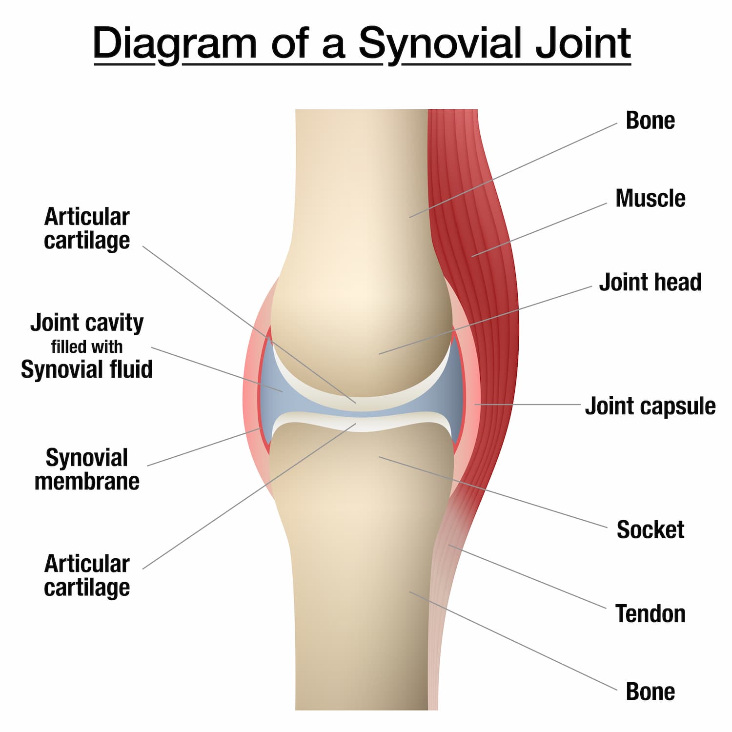
The capsule of the knee joint is a fibrous membrane containing areas of thickening that may be referred to as discreet ligaments. The anterior capsule is thin and directly anterior it is replaced by the patellar ligament. Proximally the capsule of the knee joint attaches to the femur approximately three to four finger breadths above the patella. Distally it attaches circumferentially to the tibial margin except where the popliteal tendon enters the joint through the hiatus. Posteriorily the capsule consists of vertical fibres that arise from the condyles and from the walls of the intercondylar fossa of the femur. In this region the capsule is augmented by the fibres of the oblique popliteal ligament, which is derived from the semi-membranous tendon. This broad flat band is attached proximally to the margin of the intercondylar fossa and posterior surface of the femur close to the articular margins of the condyles. The oblique popliteal ligament forms part of the floor of the popliteal fossa and the popliteal artery rests on it.
Because almost all tendons at the knee lie parallel to the bone and pull lengthwise across the joint, bursae are numerous (Fig 3, page 61, The adult knee). Bursae are potential sacs separating tendons from bones and skin. They contain a microscopic fluid layer and allow tendons to move over bones smoothly. They can become inflamed and fill with fluid to form lumps around the knee.
The suprapatellar bursa lies between the quadriceps tendon and the anterior femur. Three other bursae are associated with the patella and its ligament. The prepatellar bursae located between the skin and the anterior surface of the patella allows free motion of the skin over the patella during flexion and extension. The subcutaneous infrapatellar bursa lies between the patellar tendon and the overlying skin. Both these bursae can become inflamed during repetitive activities such as kneeling or the result of direct trauma. The deep infrapatellar bursa located between the patellar ligament and the tibial tuberosity is separated from the synovial cavity of the joint by the infrapatellar fat pad and helps to reduce friction between the patellar ligament and the tibial tuberosity.
Medially the bursa anserina lies deep to the pes anserinus tendons (sartorius, gracilis and semi tendinosis) and separates them from the tibial collateral ligament. The bursa of the semi membranosis muscle lies between the muscle and the tibia.
Posteriorily there are two large bursae associated with the medial and lateral heads of the gastrocnemius muscle. The bursa of the medial head of the gastrocnemius underlies the medial head separating it from the capsule and generally communicates with the knee joint. This bursa is the most common site of the occurrence of a Baker’s cyst.
There are numerous other bursae around the knee, many of which can communicate with the knee joint to produce swellings around the knee.
Make An Enquiry
Or contact us directly
[email protected]
0161 445 4988